17 questions

Breast surgery: augmentation, reduction and mastopexy

Common questions on Mentor and Motiva implants, incision choice and scarring, breastfeeding, capsular contracture and breast cancer screening after augmentation.

Breast surgery: augmentation, reduction and mastopexy
17
01

What is the difference between Mentor and Motiva breast implants?

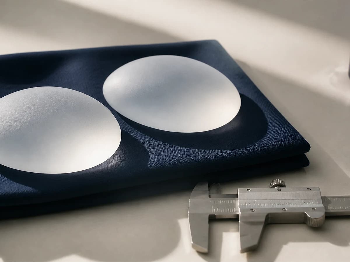
Both are American brands; Motiva manufactures in Costa Rica. Mentor is longer established with extensive long-term follow-up data, offering textured or smooth shells with cohesive gels of varying firmness. Motiva uses an ultra-smooth SmoothSilk shell and a highly elastic gel that changes shape with posture. The choice depends on your breast tissue and desired shape.

Clinical detail

The difference patients actually perceive lies in softness and how the shape changes between standing and lying. Motiva Ergonomic gel tends to settle into the lower pole when upright and redistribute when supine, closely mimicking natural breast behaviour. Mentor Gel Xtra is firmer and holds its shape more consistently, which suits patients with thin tissue who need structural support.

In patients with thin glandular tissue, an overly firm gel readily shows the implant edge at the upper pole. In those with dense tissue, a softer gel may not create the fullness sought. This is why implant selection must follow actual measurement rather than brand preference.

What influences the outcome more than implant choice is accurate breast base measurement, correct volume and profile selection, the plane of placement and pocket dissection technique. A good implant placed poorly still produces a poor result.

Clinically reviewed by Dr. Le Trung Kien, MD, Specialist Level II · Updated 12/08/2026. This information is for reference; the final indication rests with your surgeon after an in-person examination.
02

How long do breast implants last, and do they need routine replacement?

Breast implants have no fixed expiry date and do not require scheduled replacement. In practice, a proportion of patients undergo revision surgery within 10 to 15 years for reasons such as capsular contracture, implant rupture, a change in desired size, or changes in breast tissue with age and weight.

Clinical detail

The belief that implants "must be replaced after 10 years" is a common misconception. No medical guideline requires scheduled replacement in an asymptomatic patient with normal imaging.

What is recommended is regular surveillance. For silicone gel implants, international guidance advises scheduled ultrasound or MRI to detect silent rupture, since cohesive gel rupture often produces no change in breast shape and no symptoms.

The most frequent reason for revision in practice is not implant failure but change in the surrounding breast tissue: age-related ptosis, or volume change after pregnancy or weight loss. Management then usually involves mastopexy with implant exchange rather than exchange alone.

Clinically reviewed by Dr. Le Trung Kien, MD, Specialist Level II · Updated 12/08/2026. This information is for reference; the final indication rests with your surgeon after an in-person examination.
03

Can I breastfeed after breast augmentation?

Most women can breastfeed after implant-based augmentation, because the implant is placed behind the gland or behind pectoralis major without disturbing glandular tissue or the ductal system. The largest determinant is incision choice: a periareolar approach carries higher risk to the ducts than an inframammary one.

Clinical detail

If you plan future pregnancies, state this at the consultation. It directly influences incision choice and implant plane, which can be adjusted to preserve glandular function as far as possible.

This must be distinguished from breast reduction. Reduction involves excising glandular tissue and repositioning the nipple-areola complex, so its impact on lactation is considerably greater than implant augmentation.

Pregnancy and lactation alter breast volume and ptosis even with implants in place. This is why RASA often advises timing augmentation around family plans, not because of medical risk but because of the durability of the aesthetic result.

Clinically reviewed by Dr. Le Trung Kien, MD, Specialist Level II · Updated 12/08/2026. This information is for reference; the final indication rests with your surgeon after an in-person examination.
04

What is capsular contracture and how is it prevented?

The body always forms a connective tissue capsule around an implant; this is a normal response. Capsular contracture occurs when that capsule thickens and tightens, making the breast firm, distorted and sometimes painful. It is the most common late complication of implant-based augmentation.

Clinical detail

Severity is graded on the Baker scale from I to IV. Grades I and II usually require no intervention. Grades III and IV cause visible distortion or pain and require revision surgery with capsulectomy and implant exchange.

Risk-reduction measures used at RASA include strict aseptic technique, minimising implant exposure to air and skin, using an insertion sleeve, irrigating the pocket with antimicrobial solution, and meticulous haemostasis to avoid haematoma. Postoperative haematoma is one of the clearest risk factors.

No measure eliminates this risk entirely. If a previously soft breast becomes progressively firm, changes shape or rides higher than before, contact your surgeon for assessment rather than waiting.

Clinically reviewed by Dr. Le Trung Kien, MD, Specialist Level II · Updated 12/08/2026. This information is for reference; the final indication rests with your surgeon after an in-person examination.
05

Where is the breast augmentation incision placed and how does it scar?

The three common approaches are inframammary, periareolar and transaxillary. The inframammary fold is the most frequently used because it allows the most precise pocket control, with the scar hidden in the natural fold and fading over 6 to 12 months. Incision length depends on implant volume: for small implants it can be as short as 2.5cm.

Clinical detail

The periareolar scar is less conspicuous because it sits at a colour boundary, but the approach traverses glandular tissue, raising the risk of ductal disruption and of infection through contact with the ductal system. It suits patients with an adequately sized areola.

The transaxillary approach leaves no scar on the breast, but surgical exposure is more limited, pocket refinement and haemostasis are harder, and revision usually requires an additional incision.

No single approach is best in all cases. The choice depends on breast anatomy, implant type, family plans and how you weigh scar position against surgical precision.

Clinically reviewed by Dr. Le Trung Kien, MD, Specialist Level II · Updated 12/08/2026. This information is for reference; the final indication rests with your surgeon after an in-person examination.

06

Is submuscular or subglandular implant placement better?

Neither plane is universally better. Submuscular placement gives a more natural upper pole, less implant edge visibility and lower contracture rates, suiting patients with thin tissue, but is more painful and the implant can move on muscle contraction. Subglandular placement is less painful with a fuller upper pole, suiting patients with ample breast tissue.

Clinical detail

In current practice the most common choice is dual-plane placement, combining both advantages: the upper pole is covered by muscle to prevent edge visibility, while the lower pole sits directly behind the gland so the implant drapes naturally.

The upper pole pinch test is the practical determinant: if pinchable tissue thickness is under roughly 2cm, subglandular placement risks visible implant edges and should be avoided.

For patients who train the upper body heavily, animation deformity on pectoral contraction is a consideration when choosing a submuscular plane.

Clinically reviewed by Dr. Le Trung Kien, MD, Specialist Level II · Updated 12/08/2026. This information is for reference; the final indication rests with your surgeon after an in-person examination.

07

How is implant size chosen for a natural result?

Implant size is derived from breast base width, chest dimensions, tissue thickness and skin elasticity, not from a desired cc figure. An implant wider than the breast base will encroach on the axilla and the intermammary space, producing an unnatural shape and chronic tissue overstretch.

Clinical detail

The cc figure causes more confusion than anything else in augmentation consultations. The same 300cc looks entirely different on two patients with differing base width and tissue thickness. Implant width and profile are the two parameters that determine shape.

An implant too large for the supporting tissue produces late consequences: progressive skin thinning, edge visibility and rippling, early ptosis, and in some cases inferior implant migration below the fold. This is a common reason for revision after a few years.

RASA combines clinical measurement with in-bra sizer trials so you can visualise the result before deciding. The goal is the largest volume your tissue can support durably, not the largest volume that will physically fit.

Clinically reviewed by Dr. Le Trung Kien, MD, Specialist Level II · Updated 12/08/2026. This information is for reference; the final indication rests with your surgeon after an in-person examination.

08

How long does it take for breast implants to soften and settle?

During the first weeks, the breasts commonly sit higher and feel tighter and firmer than they eventually will. Most begin to soften and settle over 6 to 12 weeks, while a reasonably stable shape often takes 3 to 6 months. Timing varies with implant plane, tissue thickness, implant characteristics and individual healing.

Clinical detail

The gradual movement from an initially high position into the designed pocket is part of implant settling. The two sides may not progress at exactly the same rate because swelling, muscle tone and baseline anatomy are rarely identical.

Do not massage forcefully or try to push an implant down unless specifically instructed. Postoperative handling differs according to surgical technique and pocket condition; a manoeuvre suitable for one patient may be inappropriate for another.

Increasing redness, heat or pain, rapid one-sided swelling, wound discharge or a sudden change in implant position warrants early contact with the surgical team rather than waiting for the 3 to 6 month mark.

Clinically reviewed by Dr. Le Trung Kien, MD, Specialist Level II · Updated 12/08/2026. This information is for reference; the final indication rests with your surgeon after an in-person examination.

09

Can breast augmentation reduce nipple sensation?

Nipple sensation may temporarily decrease, become hypersensitive or feel numb after augmentation because tissue is stretched and small nerve branches are disturbed during pocket creation. Most changes improve over weeks to months, but a small proportion may persist. Risk depends on incision, implant size and individual anatomy.

Clinical detail

Sensation to the nipple-areola complex is supplied mainly by intercostal nerve branches entering from the lateral breast. Excessively wide pocket dissection or an implant that places substantial tension on the breast base increases the risk of sensory disturbance.

A periareolar incision passes closer to glandular tissue and the sensory network than an inframammary incision, so this trade-off deserves particular attention in patients who prioritise sensation and future breastfeeding.

Intermittent tingling and patchy numbness can occur as nerves recover. Loss of sensation accompanied by nipple colour change, increasing pain or a wound abnormality should be assessed promptly.

Clinically reviewed by Dr. Le Trung Kien, MD, Specialist Level II · Updated 12/08/2026. This information is for reference; the final indication rests with your surgeon after an in-person examination.

10

Can breast augmentation create the same close cleavage as a reference photo?

Cleavage is limited by sternum width, pectoral attachments, the distance between the breast bases and nipple direction. Implants can improve medial fullness but cannot safely reproduce the same narrow cleavage on every chest. Over-dissecting toward the midline risks symmastia and a deformity that is difficult to correct.

Clinical detail

Reference photographs are useful for describing a preference such as upper-pole fullness, a gentle slope or more visible cleavage. They are not a template that can be copied because chest width, tissue thickness and nipple position differ between individuals.

The surgeon selects implant diameter and profile to improve medial fullness while remaining within the breast footprint. An overly wide implant can cross toward the midline, extend into the axilla, thin the tissue and increase long-term edge visibility.

In patients with thin tissue or widely separated breast bases, adjunctive fat grafting may soften the medial transition. Its effect is limited and it cannot replace the underlying anatomy.

Clinically reviewed by Dr. Le Trung Kien, MD, Specialist Level II · Updated 12/08/2026. This information is for reference; the final indication rests with your surgeon after an in-person examination.

11

When can I sleep on my side, drive and return to the gym after augmentation?

Gentle walking should begin early, but back sleeping is commonly advised for about 2 to 4 weeks. Drive only after stopping sedating medication and when steering and emergency braking are pain-free, often after 1 to 2 weeks. Light lower-body exercise may resume earlier, while chest training and heavy lifting usually wait at least 4 to 6 weeks pending review.

Clinical detail

These are reference ranges rather than a fixed schedule for everyone. Submuscular placement, augmentation combined with mastopexy and revision surgery generally require a more cautious progression than straightforward subglandular augmentation.

During early recovery, a postoperative bra helps control swelling and limits unwanted implant movement. Do not switch to an underwired bra, increase compression or stop wearing support early without confirmation from the surgical team.

When returning to exercise, increase load gradually and stop if sharp pain, one-sided tightness or increased swelling follows a session. Review findings matter more than trying to meet a generic timeline found online.

Clinically reviewed by Dr. Le Trung Kien, MD, Specialist Level II · Updated 12/08/2026. This information is for reference; the final indication rests with your surgeon after an in-person examination.

12

Does breast reduction affect the ability to breastfeed?

It can, to a degree depending on technique. Reduction excises glandular tissue and repositions the nipple-areola complex, interrupting some ducts. Modern pedicle techniques preserve substantial function, but the ability to breastfeed afterwards cannot be guaranteed.

Clinical detail

If you still plan to have children, discuss this openly beforehand. In many cases the sensible option is deferring reduction until after your last delivery, since pregnancy also alters volume and ptosis and can undermine the result already achieved.

The full picture matters. Breast hypertrophy is not only an aesthetic concern: it causes neck and shoulder pain, back pain, bra strap grooving, inframammary intertrigo and restricted activity. For symptomatic patients the benefit usually outweighs the trade-offs, and this is a genuine medical indication.

Reduction leaves more scarring than augmentation, typically an inverted-T or vertical pattern. This trade-off must be accepted before deciding.

Clinically reviewed by Dr. Le Trung Kien, MD, Specialist Level II · Updated 12/08/2026. This information is for reference; the final indication rests with your surgeon after an in-person examination.

13

Sagging breasts: implants or a breast lift?

It depends on nipple position relative to the inframammary fold. If the nipple remains above the fold, an implant may suffice to restore volume and provide modest elevation. If the nipple sits at or below the fold, mastopexy is required to reposition it; an implant alone will simply make a ptotic breast larger.

Clinical detail

This is the most common indication error in breast surgery. Placing a large implant in a significantly ptotic breast adds load to already weakened tissue, accelerating ptosis and producing the picture of a high-riding implant with glandular tissue sliding beneath it.

A rough self-assessment: stand upright before a mirror and note nipple position relative to the inframammary fold. This is the basis of the Regnault ptosis grading surgeons use.

Where both volume loss and ptosis are present, the answer is mastopexy combined with augmentation in a single stage. This is more complex with more scarring, but it is the correct indication. Accepting additional scarring for a durable result is usually the sounder choice.

Clinically reviewed by Dr. Le Trung Kien, MD, Specialist Level II · Updated 12/08/2026. This information is for reference; the final indication rests with your surgeon after an in-person examination.

14

Can breast augmentation be done with autologous fat?

Yes, but the volume gain is limited, typically about one cup size per session. The advantages are use of your own tissue, entirely natural feel, no synthetic device, and simultaneous improvement of the donor area. The limitations are the need for adequate donor fat, partial resorption, and less predictable volume than implants.

Clinical detail

The best candidates want a moderate volume increase, have adequate abdominal or thigh fat, and value natural feel over size. Slim patients often lack sufficient donor tissue.

Breast fat grafting is also highly effective as an adjunct: masking implant edges in thin-tissue patients, correcting asymmetry, and softening the upper pole transition. This may be its most valuable application.

A screening note: breast fat grafting can produce calcifications or oil cysts, and radiologists need to know this in advance to distinguish them from malignant lesions. Always disclose a history of fat grafting when attending for mammography.

Clinically reviewed by Dr. Le Trung Kien, MD, Specialist Level II · Updated 12/08/2026. This information is for reference; the final indication rests with your surgeon after an in-person examination.

15

Does breast augmentation increase the risk of breast cancer?

Large-scale studies to date have found no evidence that breast implants increase the risk of conventional breast cancer. There is, however, a distinct rare entity called BIA-ALCL, an anaplastic large cell lymphoma associated with implants, reported predominantly with certain textured shell types.

Clinical detail

BIA-ALCL is a disease of the periprosthetic capsule, not of breast glandular tissue. Incidence is very low and most cases detected early are cured by en bloc capsulectomy and implant removal. The warning sign is unexplained breast swelling after years of stability, or a new mass.

Following the BIA-ALCL data, the field has shifted substantially towards smooth or nanotextured shells. Implant selection now takes this into account.

One point deserves emphasis: having implants does not exempt you from routine breast cancer screening. Self-examination, ultrasound and age-appropriate mammography schedules remain the same as for anyone else.

Clinically reviewed by Dr. Le Trung Kien, MD, Specialist Level II · Updated 12/08/2026. This information is for reference; the final indication rests with your surgeon after an in-person examination.

16

Can I still have a screening mammogram after breast implants?

Yes, but inform the imaging centre in advance that you have implants. The radiographer will add Eklund displacement views, pushing the implant back and compressing the glandular tissue separately so that areas obscured by the implant can be visualised. Ultrasound and MRI are also commonly used alongside.

Clinical detail

Implants obscure part of the glandular tissue on standard mammographic views, which is why the additional projections are necessary rather than optional. Submuscular placement obscures less than subglandular.

Screening retains its value when performed with the correct technique. What must be avoided is skipping screening in the belief that implants preclude imaging, a misconception with serious consequences.

Keep the implant information card provided by your surgical facility, recording manufacturer, shell type, volume and lot number. This is useful both for radiologists and for any future intervention.

Clinically reviewed by Dr. Le Trung Kien, MD, Specialist Level II · Updated 12/08/2026. This information is for reference; the final indication rests with your surgeon after an in-person examination.

17

When is breast implant revision surgery needed?

Common indications include Baker grade III to IV capsular contracture causing firmness or distortion, implant rupture, malposition or asymmetry, edge visibility and rippling in thin-tissue patients, progressive ptosis over time, and a wish to change size from the original result.

Clinical detail

Revision is generally more complex than primary surgery because it involves scarred tissue, an established pocket and altered anatomy. Preoperative assessment therefore needs to be more thorough, sometimes including MRI to evaluate the implant and capsule.

For thin-tissue patients with visible edges, the solution is usually not implant exchange alone but adding coverage: peripheral fat grafting, or converting from a subglandular to a submuscular plane.

RASA accepts assessment of cases operated elsewhere. If you still have your implant details and the original operative note, bring them; this makes planning considerably more accurate.

Clinically reviewed by Dr. Le Trung Kien, MD, Specialist Level II · Updated 12/08/2026. This information is for reference; the final indication rests with your surgeon after an in-person examination.

Haven’t found the answer you need?

The first consultation at RASA is free and conducted personally by Dr. Le Trung Kien. You are not asked to decide during the consultation.

Zalo
Care team support