Lecture note
Ultrasound is not limited to finding a vessel or a filler deposit. Its greater value lies in reading the face by tissue plane, documenting probe orientation and correlating images with history, examination and prior technique.
1. Scope of the module
The Morpheus8 & Clarius workshop was held in Hanoi on 20 July 2026. Dr. Le Trung Kien attended as an invited guest, documented the program and later organized the facial-ultrasound material for RASA Surgical Education. RASA was not an organizer or co-organizer of the event.
The lesson moves from image formation, presets and scanning modes to tissue-plane recognition, vascular mapping and the interpretation of commonly encountered injectable materials. Its purpose is not to turn one echo pattern into a definitive diagnosis, but to build a repeatable observation process.
- Read the image by anatomical plane rather than searching only for an isolated abnormality.
- Standardize probe orientation, pictogram and saved-image position for later comparison.
- Combine B-mode with Color or Power Doppler when the clinical question involves flow.
- Correlate imaging with product type, injection technique, timing and tissue response.
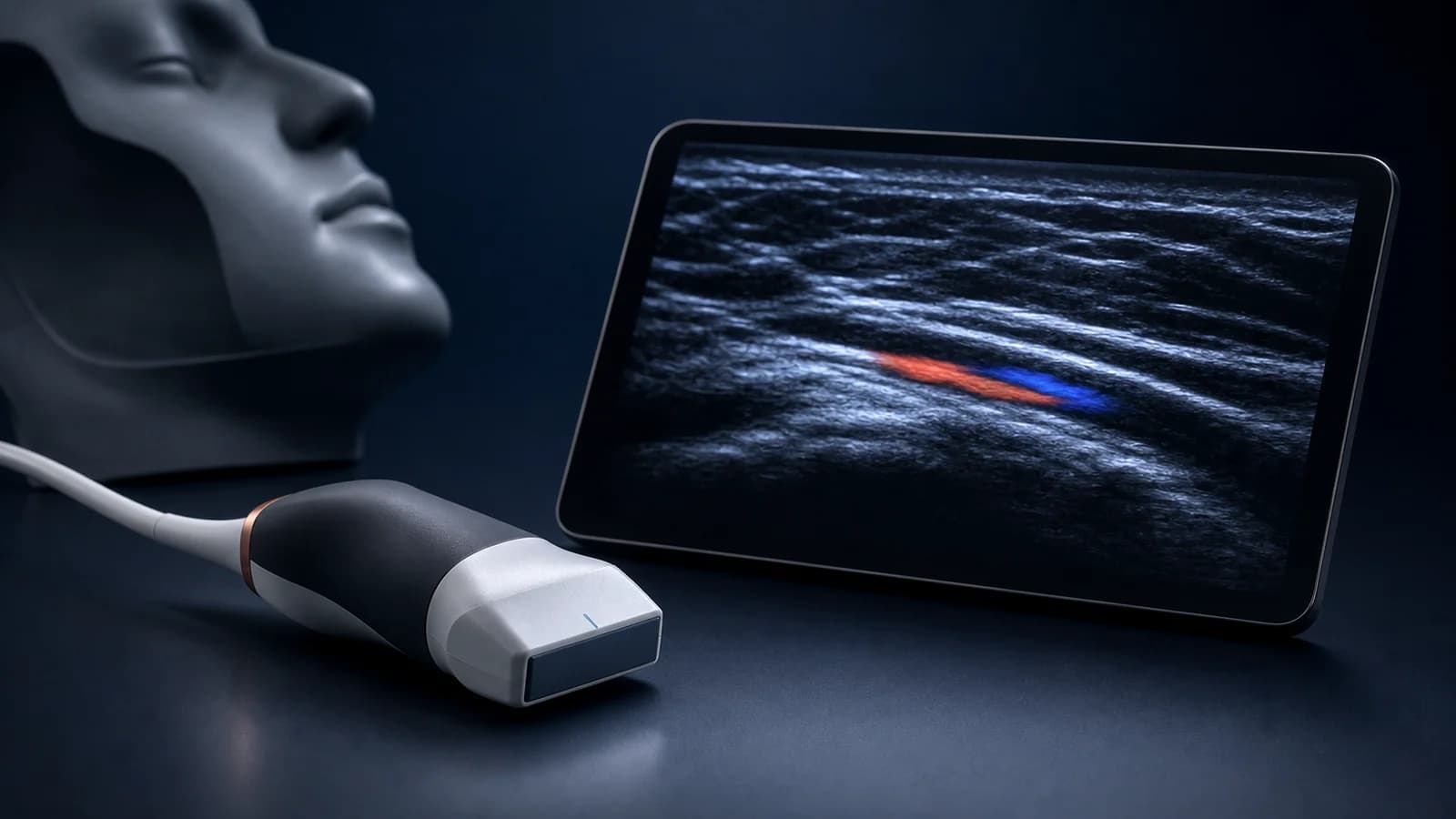
2. Why high frequency suits superficial facial structures
The core principle is the trade-off between resolution and penetration. Higher frequency can better separate superficial structures such as skin, subcutaneous tissue, fascia, muscle and the shallow spaces relevant to facial aesthetics. The trade-off is reduced penetration compared with lower-frequency probes used for deeper organs.
Image quality also depends on preset, depth, gain, focus, probe pressure, gel and scan direction. A reliable examination therefore starts by optimizing the acoustic window before interpreting echogenicity.
B-mode provides the structural foundation. Color and Power Doppler add flow information, with Power Doppler often useful for smaller or slower signals. The operator should move between modes according to the question rather than expecting one mode to answer everything.
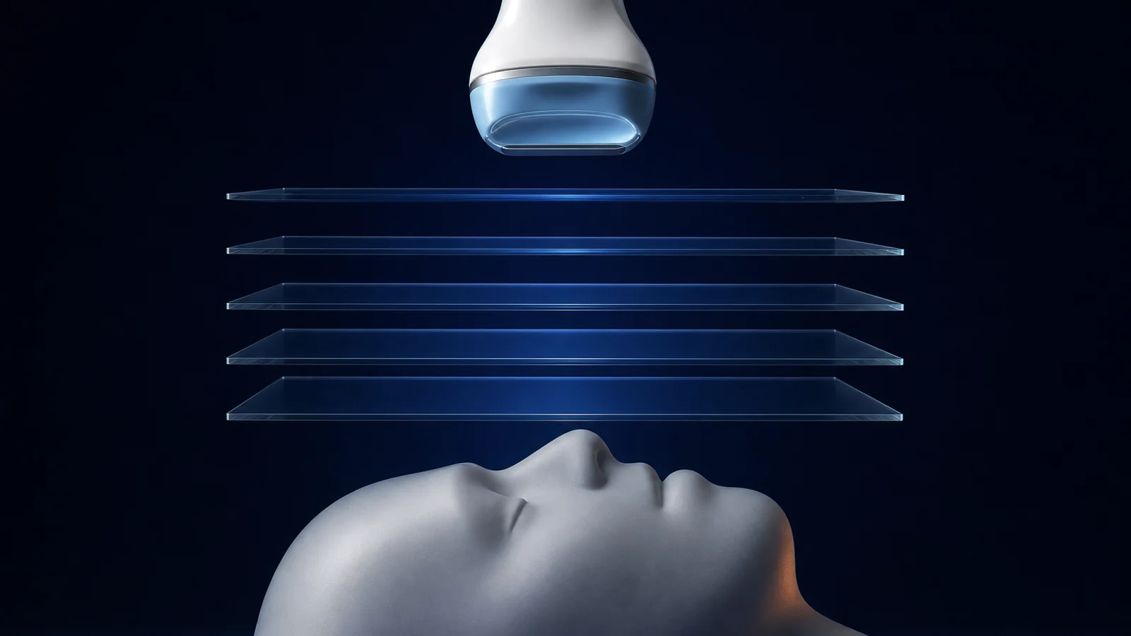
3. Standardizing presets, notch and pictogram
Before scanning, the operator selects a region-appropriate preset for the forehead, temple, cheek, lip or infraorbital area. A preset is a starting point for depth, gain and processing, not a substitute for patient-specific adjustment.
The probe orientation marker should follow a consistent convention. When an image is saved, the pictogram should record probe position and scan direction. Without this information, a visually clear image may still be difficult to compare, communicate or reproduce.
A minimum documentation set should include B-mode, anatomical location, probe direction, depth, region of interest and Doppler when the question involves vessels. This reduces the risk of interpreting a single disconnected slice.
4. Reading the face by plane and region
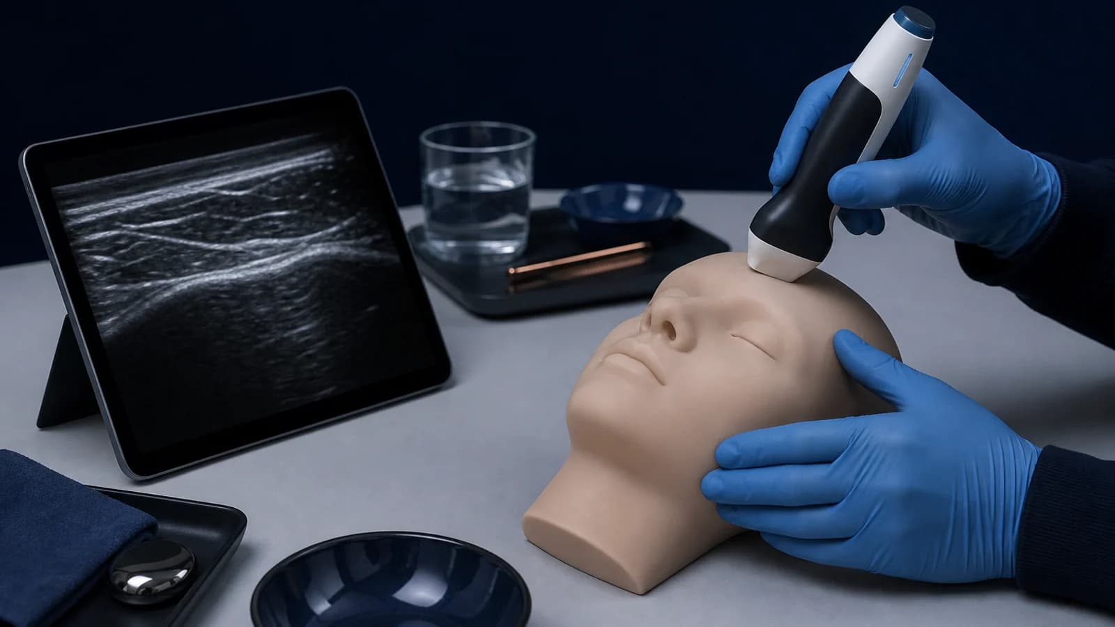
The practical session begins with the temple, where thin layers and the relationship among skin, subcutaneous tissue, superficial temporal fascia, fat spaces, deep temporal fascia, temporalis muscle and periosteum demand careful orientation. The operator first establishes the layer sequence, then searches for vessels or injected material within the correct plane.
The periocular region, forehead, cheek, masseter, parotid area and perioral region demonstrate how probe direction changes with anatomy. Stable reference structures such as bone, muscle or gland help define the relative position of vessels and prior interventions.
Ultrasound does not make anatomy simple. A bright band may represent fascia, a fibrous septum or an interface; a dark area may represent fluid, material, vessel or normal tissue depending on context. Plane-based scanning narrows the possibilities before interpretation.
5. Injectable materials: interpret signs rather than guessing a product
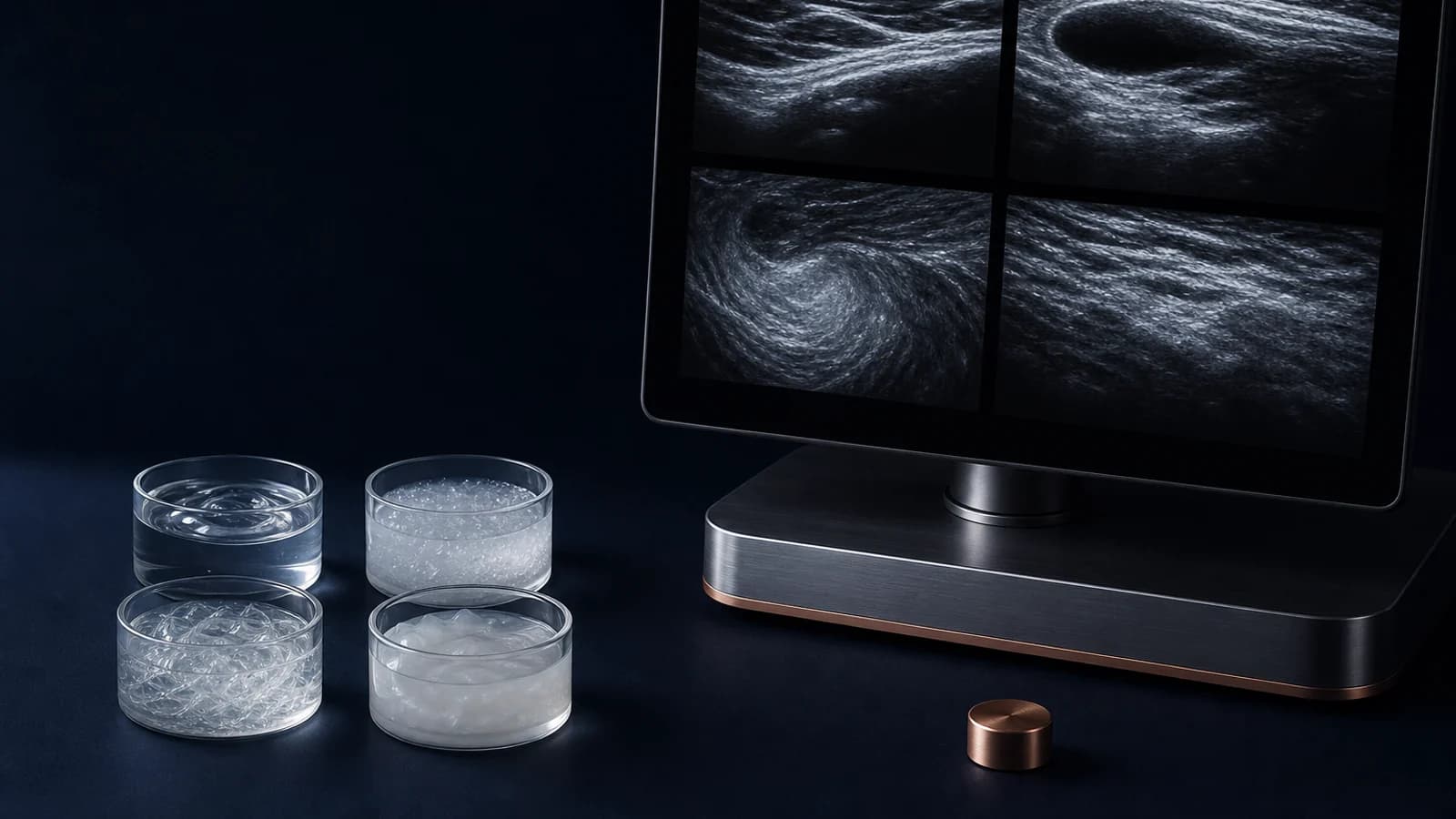
The lecture discusses HA, CaHA, PCL, PLLA and silicone. Each group may produce suggestive patterns, but appearance is not fixed. Concentration, dilution, bolus versus droplet placement, injection plane, elapsed time and tissue response can all change the image.
HA may appear relatively hypoechoic or anechoic, sometimes with defined margins and posterior enhancement. CaHA may produce bright echoes, shadowing or a different pattern after dilution and over time. Biostimulatory materials such as PCL or PLLA may appear heterogeneous with scattered bright echoes. Silicone can create artifacts and obscure deeper structures.
These descriptions are directional only. A material should not be identified from a single still image. Product history, timing, location, technique, subsequent procedures and current symptoms remain essential.
6. Doppler and vascular mapping
When the clinical question involves a vessel, Doppler adds information beyond B-mode. Color Doppler displays flow direction and distribution in a region of interest, while Power Doppler may support detection of weaker signals. Both are sensitive to scan angle, probe pressure, velocity settings and motion.
Failure to display flow does not prove that no vessel is present. Pressure may collapse a small vessel, settings may miss slow flow or the slice may not follow the vessel axis. Scanning from more than one direction and reducing pressure are important before reaching a conclusion.
Vascular mapping is only one component of safe practice. Sterility, anatomical knowledge, plane selection, instrument control and preparedness for complications remain mandatory.
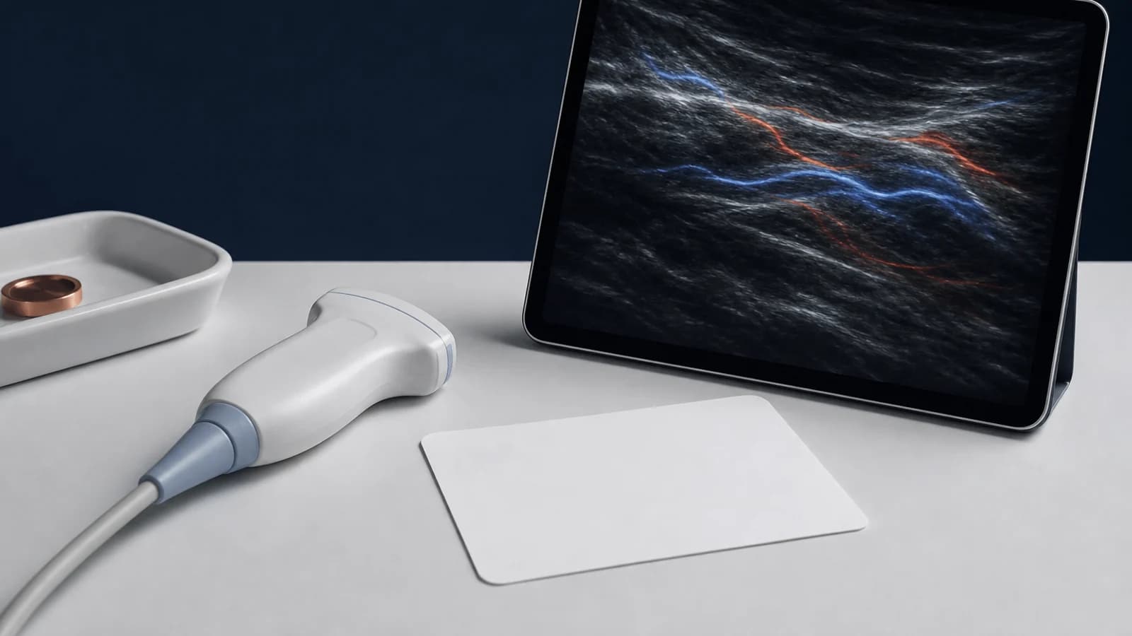
7. Clinical applications and interpretive limits
Ultrasound may help define the plane of previously injected material, distinguish focal from diffuse distribution, assess its relationship to vessels and follow changes over time. It can also help explain persistent fullness, irregularity or symptoms long after an earlier intervention.
Ultrasound does not determine treatment on its own. Image quality is operator-dependent, material patterns overlap and inflammation, fibrosis or repeated procedures can make the appearance complex. A responsible report separates direct observations from inference and states the level of certainty.
This material is intended for professional education. It does not replace direct assessment, official device instructions, supervised hands-on training or a medical facility complication protocol.
8. Course structure in RASA Surgical Portal
The course is organized into two lessons: theory covering imaging principles, tissue planes and injectable-material patterns; and a practical lesson covering device setup, probe orientation and regional facial scanning. The complete written material is available in the portal during this stage.
Video and subtitle tracks will be attached to the corresponding lessons after the release versions are completed. Separating the learning content from the media pipeline keeps the academic material available without depending on video processing progress.